Newly seen age-related macular degeneration genetic markers unveiled
Thanks to the identification of new genetic markers for age-related macular degeneration (AMD), improved detection and treatment of the incurable progressive eye disease is inching closer.
Researchers from the Garvan Institute of Medical Research (New South Wales, Australia) the University of Melbourne (Australia), the Menzies Institute for Medical Research at the University of Tasmania (Hobart, Tasmania) and the Centre for Eye Research Australia (Melbourne, Australia), created models of diseased eye cells using reprogrammed stem cells and then examined DNA, RNA and proteins to identify the genetic markers.
“We’ve tested the way that differences in people’s genes impact the cells involved in age-related macular degeneration. At the smallest scale we’ve narrowed down specific types of cells to pinpoint the genetic markers of this disease,” explains co-lead of the study Professor Joseph Powell, Pillar Director of Cellular Science at Garvan. “This is the basis of precision medicine, where we can then look at what therapeutics might be most effective for a person’s genetic profile of disease.”
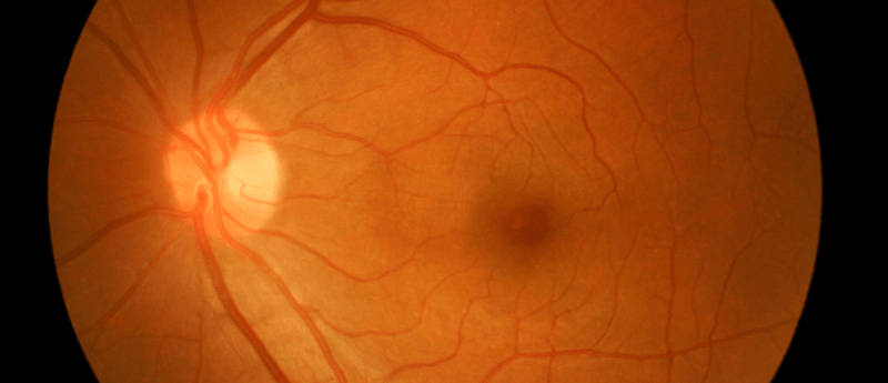
AMD is described as the gradual degeneration of a region in the middle of the retina, located behind the eye, known as the macular. With time, the condition can progress to visual impairment or the complete loss of vision. The number of Americans who suffer from age-related macular degeneration is estimated to be 11 million; by 2050, this figure is projected to double to 22 million.
Although the fundamental causes of the decline are yet unknown, environmental and genetic factors play a part. Age, family history and smoking are all risk factors.
Nature Communications recently published an article detailing the discovery of the genetic markers of the disease. 79 subjects, both with and without geographic atrophy, the late stage of AMD, had their skin samples collected by the researchers. Their skin cells were reprogrammed and reverted to become induced pluripotent stem cells, which were subsequently directed by molecular signals to become retinal pigment epithelium cells. These retinal pigment epithelium cells are the cells that are impaired by AMD, they coat the back of the retina and are crucial to the retina’s health and functionality. The loss of vision in AMD is brought on by the degeneration of photoreceptors, which are light-sensing neurons in the retina that relay visual information to the brain.
439 molecular fingerprints associated to AMD were discovered by analysis of 127,600 cells, with 43 of them being possible novel gene variants. The implicated key pathways were identified and consequently examined within the cells, exposing the disparities in the mitochondria between healthy and AMD cells. This resulted in mitochondrial proteins becoming viable targets to stop or modulate the progression of AMD.
Additionally, the molecular markers can now be utilized for the screening of treatments using patient-specific cells in a dish. “Ultimately, we are interested in matching the genetic profile of a patient to the best drug for that patient. We need to test how they work in cells relevant to the disease,” explains co-lead of the study Professor Alice Pébay, from the University of Melbourne.
In their previous work, Professor Powell and co-lead authors Professor Pébay and Professor Alex Hewitt from the Menzies Institute for Medical Research in Tasmania and the Centre for Eye Research Australia, have discovered genetic markers of glaucoma, a similar degenerative eye disease. To generate results, they utilized stem cell models of both the retina and optic nerve. The researchers are currently focusing their attention on the genetic causes of Parkinson’s and cardiovascular disorders.
Source: https://www.nature.com/articles/s41467-022-31707-4